BPPV Visualization with a 3D Model for Study and Teaching (swipe up)
Ewald’s Law in BPPV
Ewald’s First Law
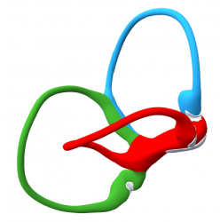
Ewald’s First Law states eye movements are in the plane of the canal being stimulated. This video shows eye movements associated with each of the 6 canals. The eye always moves in the plane of the canal stimulated or inhibited and rotates about the axis of the canal shown transposed onto the eye. Slow phase eye movements are indicated by red arrows. Fast phase eye movements are indicated by Yellow arrows. These directions of eye drift and correction correspond to the eye movements observed in BPPV when generated from these canals in the Dix Hallpike position.
Ewald’s First Law states eye movements are in the plane of the canal being stimulated. This video shows eye movements associated with the right horizontal canal. The axis of the right horizontal canal is shown transposed onto the eye- Red Rod. Slow phase eye movements are indicated by red arrows. Fast phase eye movements are indicated by Yellow arrows.
Ewald’s First Law states eye movements are in the plane of the canal being stimulated. This video shows eye movements associated with the right superior canal. The axis of the right superior canal is shown transposed onto the eye- Blue Rod. Slow phase eye movements are indicated by red arrows. Fast phase eye movements are indicated by Yellow arrows.
Ewald’s First Law states eye movements are in the plane of the canal being stimulated. This video shows eye movements associated with the right posterior canal. The axis of the right posterior canal is shown transposed onto the eye- Green Rod. Slow phase eye movements are indicated by red arrows. Fast phase eye movements are indicated by Yellow arrows.
Ewald’s First Law states eye movements are in the plane of the canal being stimulated. This video shows horizontal eye movements associated with the left superior canal. The axis of the left superior canal is shown transposed onto the eye-Blue Rod.
The characteristic Down-beating and rotary nystagmus seen in Superior canal BPPV is seen-Yellow Arrow.
Ewald’s First Law states eye movements are in the plane of the canal being stimulated. This video shows horizontal eye movements associated with the left posterior canal. The axis of the left posterior canal is shown transposed onto the eye-Green Rod. The characteristic upbeating and rotary nystagmus seen in BPPV is seen-Yellow Arrow. Red Arrow- Slow Phase/ Yellow Arrow-Fast Phase.
Ewald’s First Law states eye movements are in the plane of the canal being stimulated. This video shows horizontal eye movements associated with the left horizontal canal.
Eye Movement in Left Posterior Canal BPPV. This video demonstrates the position of the left labyrinth in the left Dix-Hallpike position. The axis of the left posterior canal is transposed onto the eye-Green Bar. The otolith mass falls around the circumference of the canal as indicated by the Small Red Arrow. The eye moves in the same direction as the otolith mass and in the Plane of the left posterior canal-Large Red Arrow. The fast phase is also in the plane of the canal- Large Yellow Arrow- and results in the upbeating geotropic rotary nystagmus characteristic of posterior can BPPV. To see the effect of gaze direction on this nystagmus see the related videos: Eye Movement in Left Posterior Canal BPPV with Left Gaze, and Eye Movement in Left Posterior Canal BPPV with Right Gaze.
Eye Movement in Left Posterior Canal BPPV with Left Gaze. This video demonstrates the position of the left labyrinth in the left Dix-Hallpike position. The axis of the left posterior canal is transposed onto the eye-Green Bar. The otolith mass falls around the circumference of the canal as indicated by the Small Red Arrow. The eye moves in the same direction as the otolith mass and in the Plane of the left posterior canal-Large Red Arrow. The fast phase is also in the plane of the canal- Large Yellow Arrow. With Left Gaze, into the axis of the canal-Green Rod- the pure rotary component of the nystagmus can be seen. Clinically this is useful when the origin of the nystagmus is not clear. Gaze to the Right will show a pure upbeating nystagmus. See the video: Eye Movement in Left Posterior Canal BPPV with Right Gaze To see the effect of gaze direction on this nystagmus see the related videos: Eye Movement in Left Posterior Canal BPPV, Eye Movement in Left Posterior Canal BPPV with Left Gaze and Eye Movement in Left Posterior Canal BPPV with Right Gaze.
Eye Movement in Left Posterior Canal BPPV With Right Gaze. This video demonstrates the position of the left labyrinth in the left Dix-Hallpike position. The axis of the left posterior canal is transposed onto the eye-Green Bar. The otolith mass falls around the circumference of the canal as indicated by the Small Red Arrow. The eye moves in the same direction as the otolith mass and in the Plane of the left posterior canal-Large Red Arrow. The fast phase is also in the plane of the canal- Large Yellow Arrow. With Left Gaze, into the axis of the canal-Green Rod- the pure rotary component of the nystagmus can be seen. With Right Gaze(SHOWN) a pure upbeating nystagmus results. Gaze Separation to show the individual components of nystagmus is useful when the origin of the nystagmus is not clear. To see the effect of gaze direction on this nystagmus see the related videos: Eye Movement in Left Posterior Canal BPPV, Eye Movement in Left Posterior Canal BPPV with Left Gaze, and Eye Movement in Left Posterior Canal BPPV with Right Gaze.
Ewald’s Second Law
The second law states that excitation of any canal creates a greater response than inhibition. This is explained physiologically in the image below. The resting discharge of the ampulla is near 170 beats per second. With maximal stimulation the discharge rate can be increased by 330 beats per second to 500. The output can be inhibited by only 170 beats per second to zero.
Ewald’s Third Law
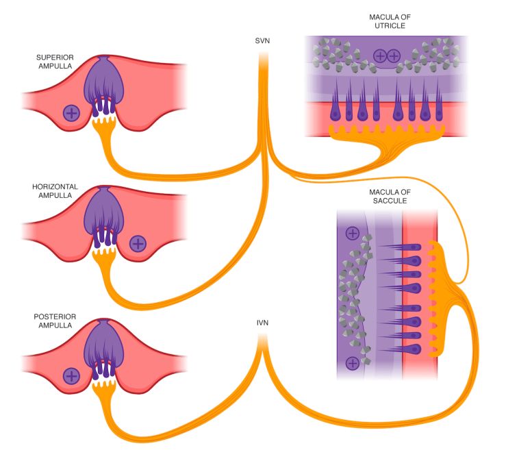
The third law clarifies the direction of polarization of the cristae and states that ampullopetal flow creates a stronger response in the lateral canal, and ampullofugal flow creates the strongest response in the anterior and posterior canals. This is summarized in the illustration below.
This illustration also clarifies the innervations of the vestibular organs, and the striolar polarizations of the maculae of the utricle and saccule. cVEMP tests the inferior vestibular nerve via the saccule. oVEMP tests the superior vestibular nerve via the utricle.